|
The Spine
Although technically a part of the skeletal
system, even on a physical level the spine is the center of
yogic anatomy, and therefore deserves special mention. In
addition to simply supporting the head, the spine is a bony,
protective canal for the spinal cord, the great central
trunk line of nerves that issues out from the brain toward
every part of the body (It's like the cable in "cable
TV.")
Vertebrae, Discs and Nerves
The spine is made of a series of
vertebrae, which are roughly cylindrical building blocks
of bone, each with a bony "cage" on its posterior to protect
the spinal cord as it runs the length of the spine. The shapes
of the vertebrae vary depending on where they are located in
the spine: cervical region, thoracic region or lumbar region.
The most prominent features of the "cage"
behind the vertebral body are a spinous process (which
sticks straight out behind you; the "bumps" on your back) and
two transverse processes (which stick out to the side
and slightly behind). In addition to helping protect the
spinal cord, the processes are important attachment points for
muscles and ligaments, as we'll soon see.
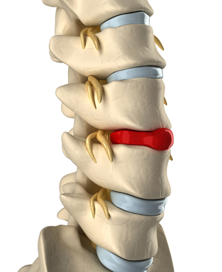
Each pair of vertebrae is separated by a
small, but very important, shock-absorbing cushion called a
disc. Discs are made of fibro cartilage, which means that
they're firm, but will yield with pressure. The center of each
disc is filled with a gel-like substance. (Image: The soles of
some running shoes are made of two layers of tough rubber,
with gel in-between for shock absorption. All movement of the
spine occurs at these discs and is limited by two
factors:
• The degree of firmness or squashiness of the
discs.
• The ligaments that span the discs, and
connect each vertebra with the vertebrae above and below
it.
At each disc joint, the vertebrae can bend
forward, backward and sideways; they also can rotate just a
little bit. The overall movement of the spine is the sum of
all these little movements. Such movements cause the gel to
move within the discs: backward during a Forward bend, forward
during a backward bend, and to one side when bending to the
opposite side.
Young discs tend to be very squashy; their
centers are almost fluid. However, discs tend to get thinner
as we age, and if we do not exercise the disc joints
adequately, the discs will dry out and shrink into hard little
plates. In this condition they limit motion severely, and they
can no longer serve as shock-absorbers for the
vertebrae.
In addition to the disc joints between the
bodies of the vertebrae, each pair of vertebrae also touch at
two other points in the bony "cage" behind the body of the
vertebrae. These points are called the facet joints,
and it is these that your chiropractor adjusts. The facet
joints have different configurations in the cervical, thoracic
and lumbar regions of the spine (see below), resulting in
different movement capabilities in each section.
A pair of nerves exit the spinal
column between each pair of vertebrae at the level of the
disc. One nerve exits to the left and one to the right, each
through a hole called a spinal foramen. The top half of
the hole is formed by a "cutout" area in the top vertebra, the
bottom half by a cutout area in the bottom one. These nerves
are the communication channels between the brain and every
cell in the body, and we'll discuss them when we study the
nervous system.
When two vertebrae are
misaligned, their two spinal foramina may no longer be nice,
rounded holes. The nerves may then be pinched where they exit
the spinal cord. This hurts.
In fact, even just rounding the spine
excessively can alter these holes and thus impinge upon the
nerves. Nerves can also be impinged upon by slipped discs
(i.e., discs that scoot backward and rest against a nerve),
hen-dated discs (i.e., discs that abnormally bulge backward
against a nerve), and ruptured discs (leaking gel;). You may
sometimes hear "herniated" used to mean,
"ruptured."
The Five Regions of the
Spine
The spine consists of 26 pieces, which are
usually divided into five sections or regions. From top to
bottom, these are:
• Cervical spine
7
vertebrae. The skull rests on the topmost vertebra, called
the atlas. The atlas and the second cervical vertebra
(the axis) have special modifications to support the movements
of the head. These vertebrae are usually referred to as C1 (or
atlas, the top vertebra), C2 (or axis, the second vertebra),
and so on down to C7, which is the bump you can feel at the
base of your neck.
• Thoracic spine
12
vertebrae, usually referred to as T1 (top) through T12
(bottom). These are the vertebrae to which the ribs (thoracic
cage) attach.
• Lumbar spine
5
vertebrae, usually referred to as LI (top) through L5
(bottom).
• Sacral spine, or
sacrum 5 vertebrae that are fused into a single unit in
adults.
• Coccyx (tailbone) 3
or 4 tiny vertebrae that are fused into a single unit at the
base of the spine.
© 1997-2000 Ananda Church of
Self-Realization
|